


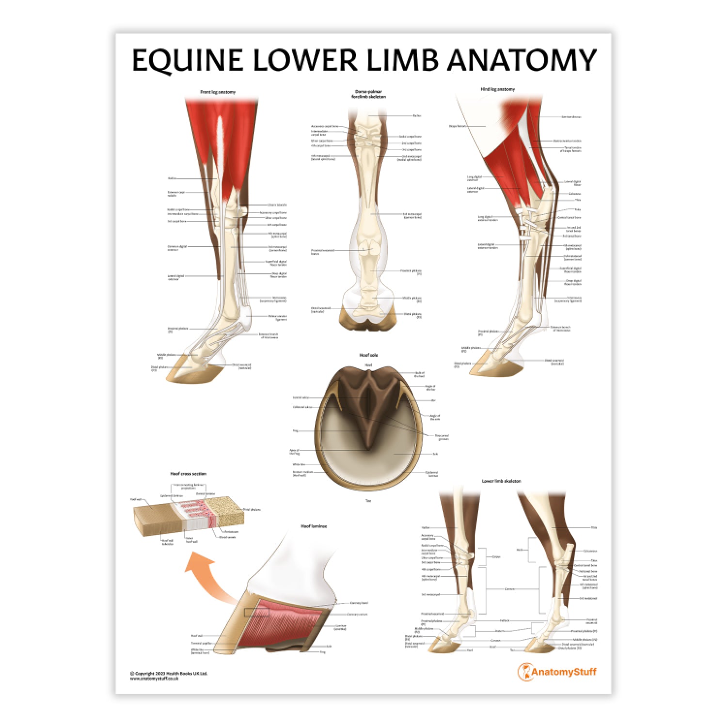
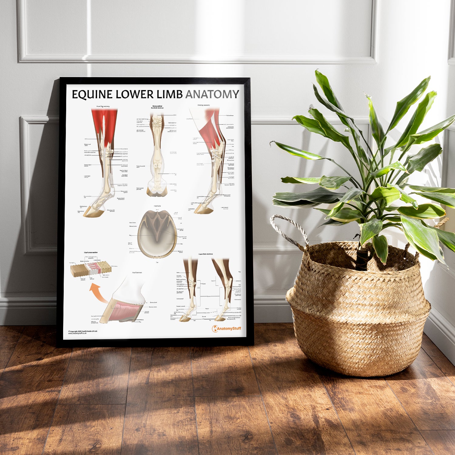
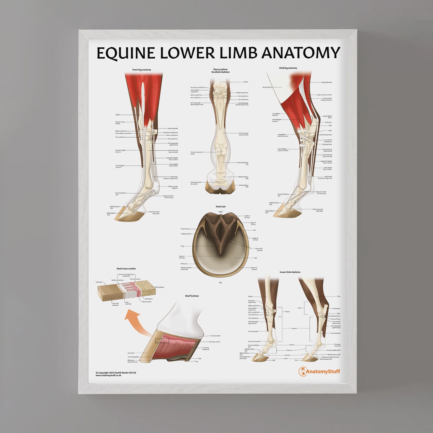

The Equine Lower Limb Anatomy Laminated Chart/Poster serves as a valuable resource for veterinary professionals or students. This meticulously crafted chart offers comprehensive views of the lower limb anatomy of horses with exceptional clarity. It is an ideal educational tool for undergraduate studies and can also be utilised as a visual aid in clinical settings.
Developed by a skilled medical illustrator and exclusive to AnatomyStuff, the Equine Lower Limb Anatomy Chart provides detailed insights into the following:
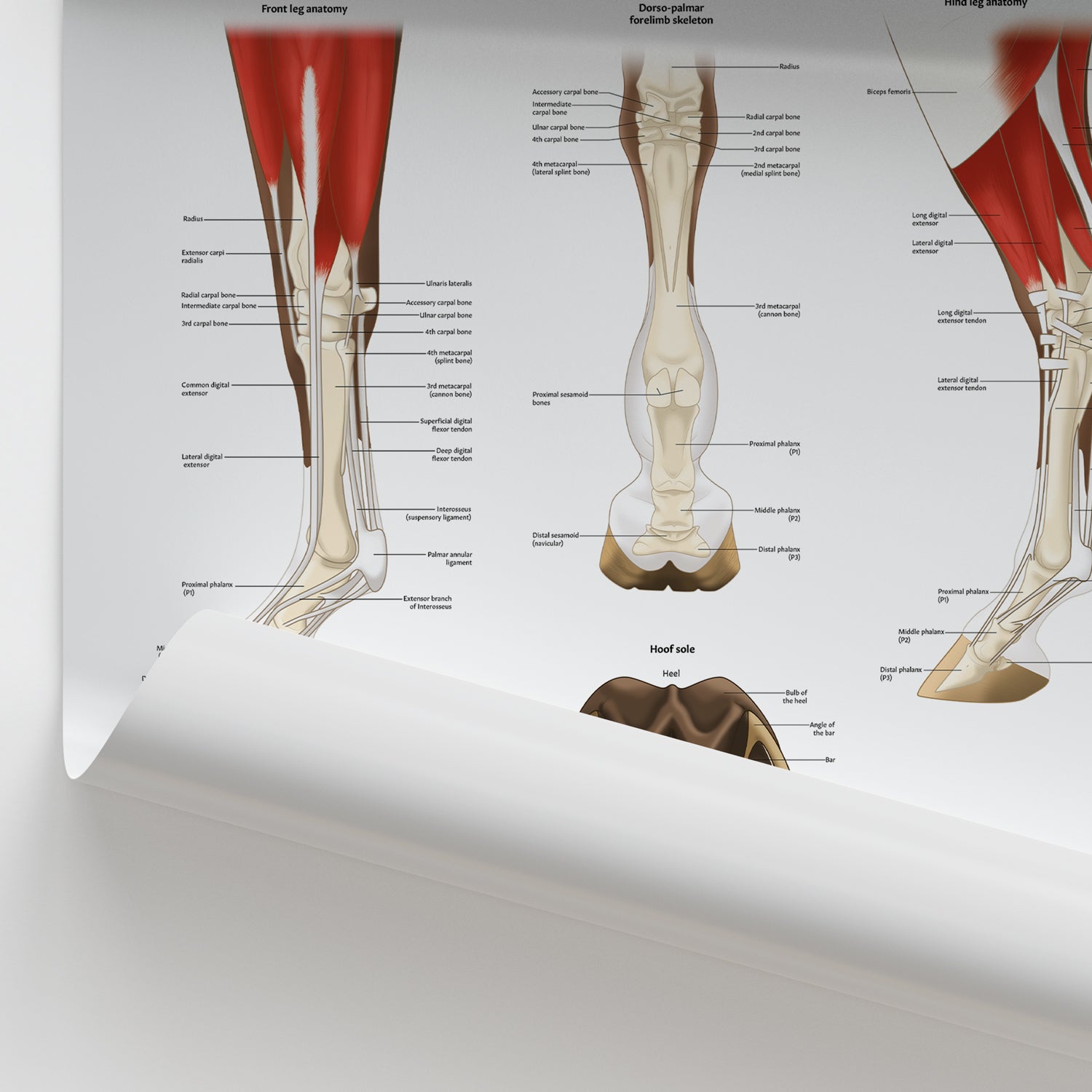
- Detailed equine front leg anatomy
- Skeletal framework of the forelimb from both dorsal and palmar views
- Comprehensive anatomy of the equine hind leg
- Structure of the hoof sole
- Cross-sectional view of the equine hoof
- Anatomy of the laminae structure of the hoof
- Skeletal anatomy of the lower limb
Our collection offers a range of display options to meet your needs:
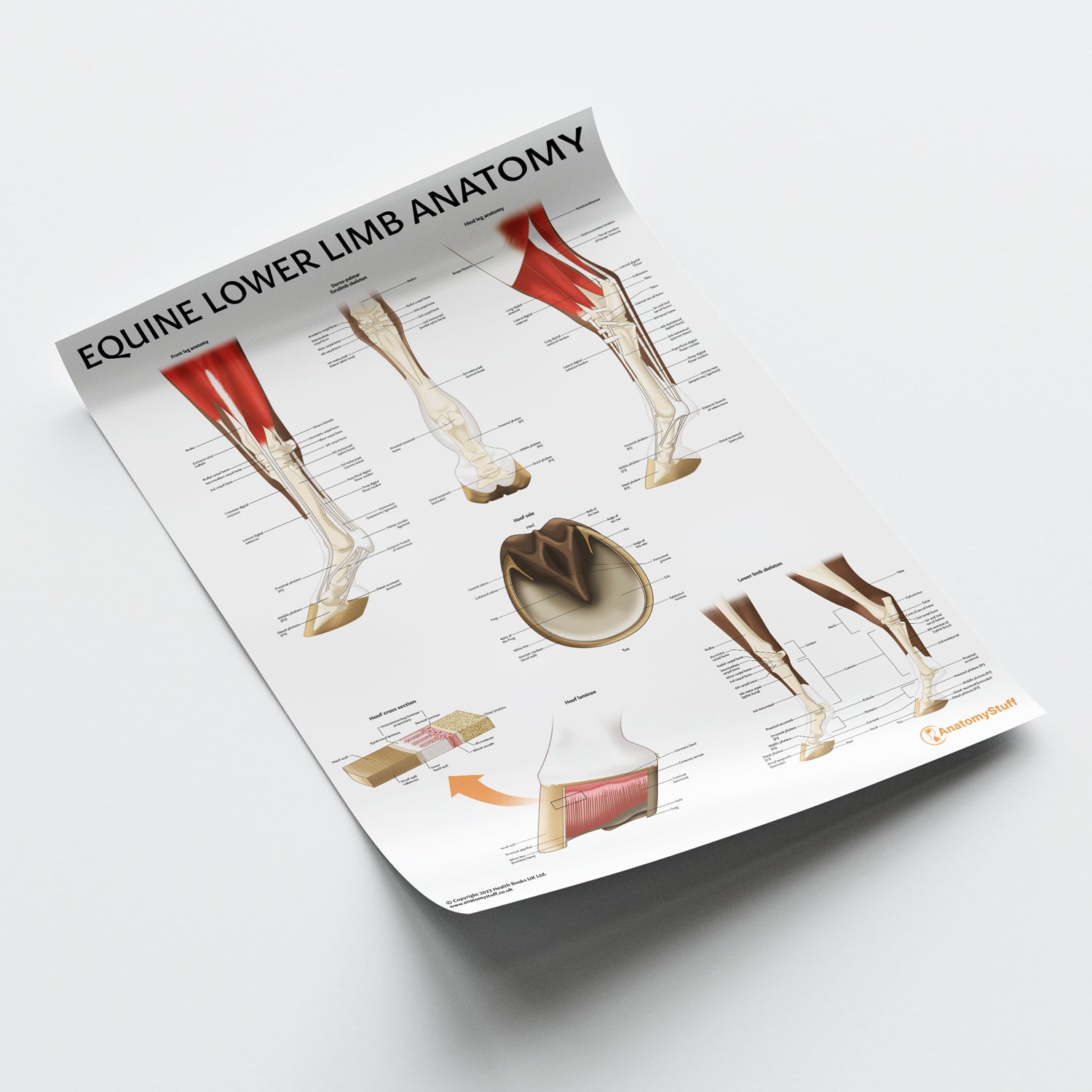
- Laminated Poster (50 x 65 cm): Ideal for clinic displays, featuring clearly labelled anatomical details. The durable lamination allows for annotation with washable markers (not supplied).
- Classic Semi-Glossy Prints: 45 x 60 cm, 60 x 80 cm, and 70 x 100 cm sizes. The semi-glossy finish enhances colours with a subtle shine, adding vibrancy to any setting.
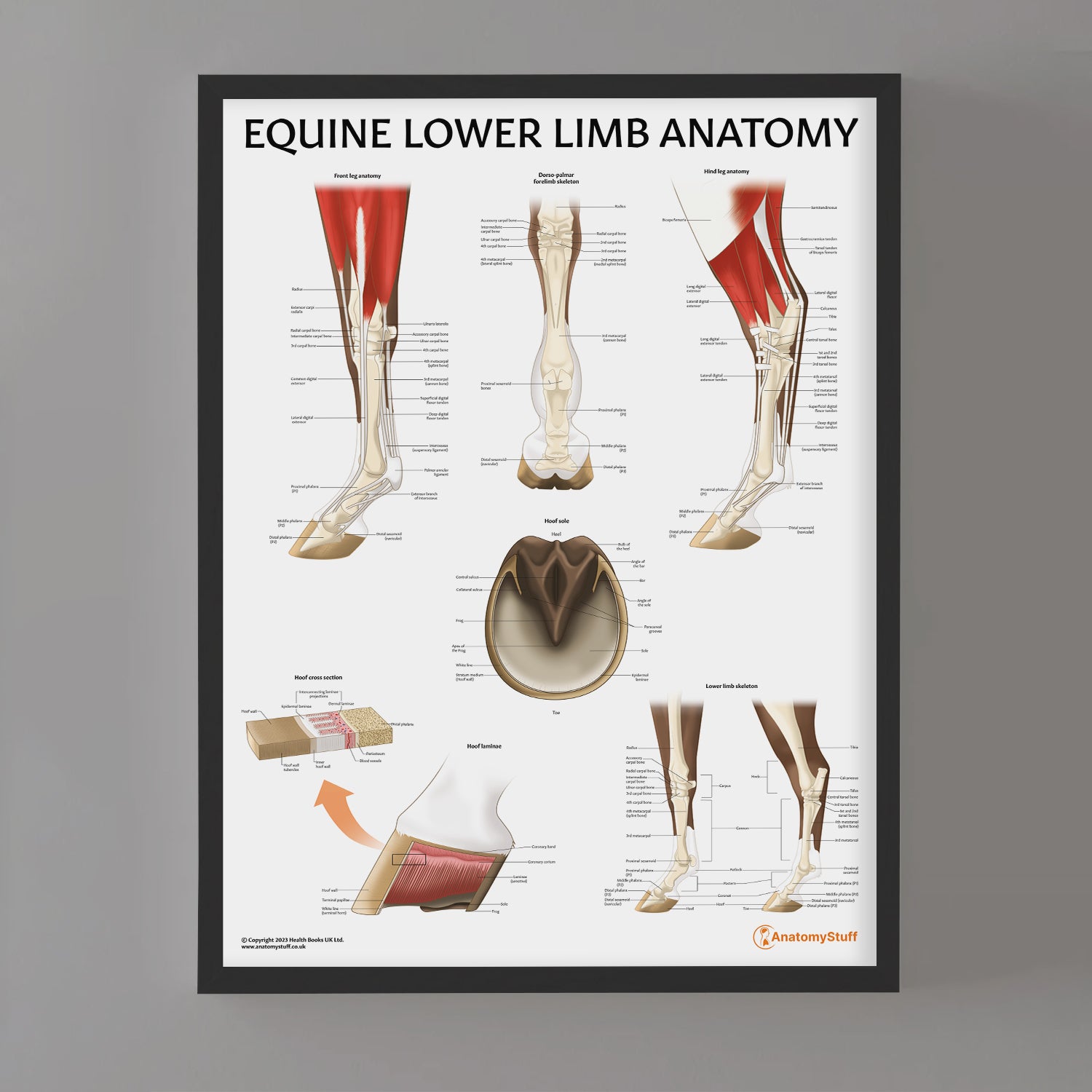
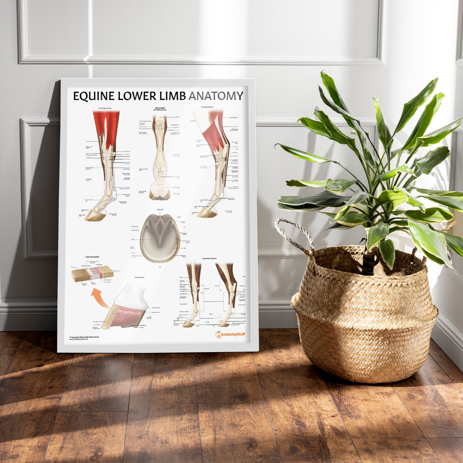
- Framed Prints: Offered in black or white frames and sizes 45 x 60 cm, 60 x 80 cm, and 70 x 100 cm. Features 170 gsm matte paper with a smooth, non-reflective finish. Ready-to-hang with a durable pine wood frame.
