


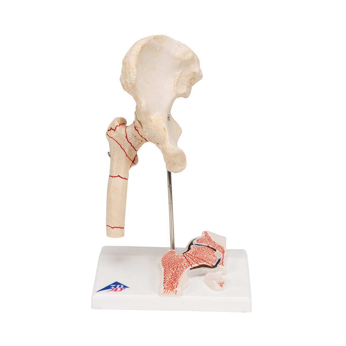
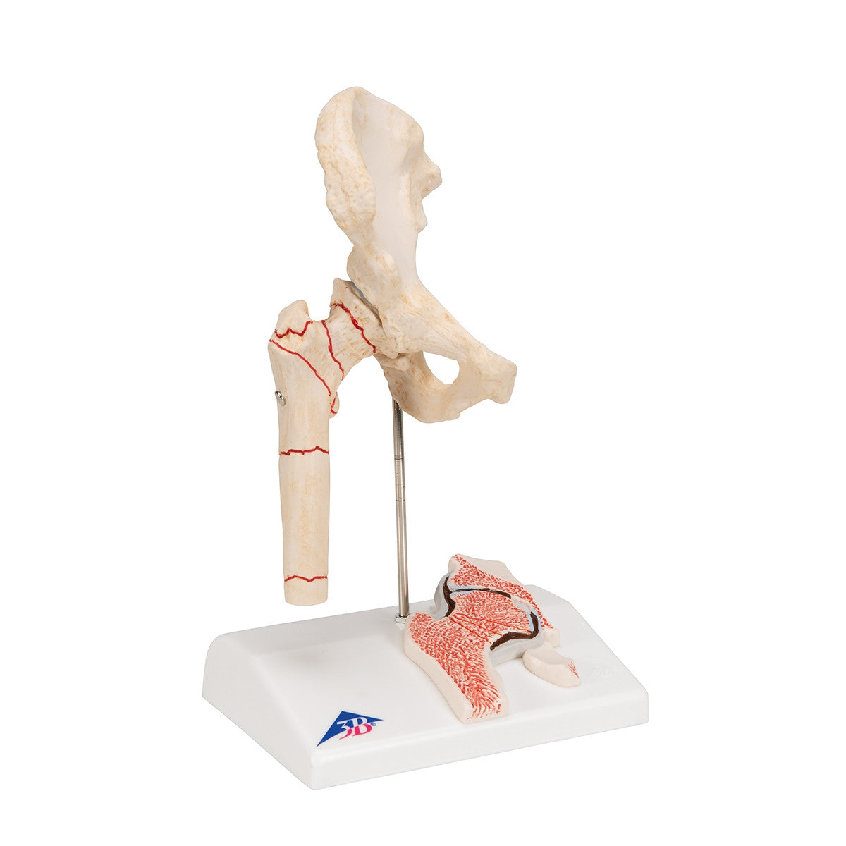
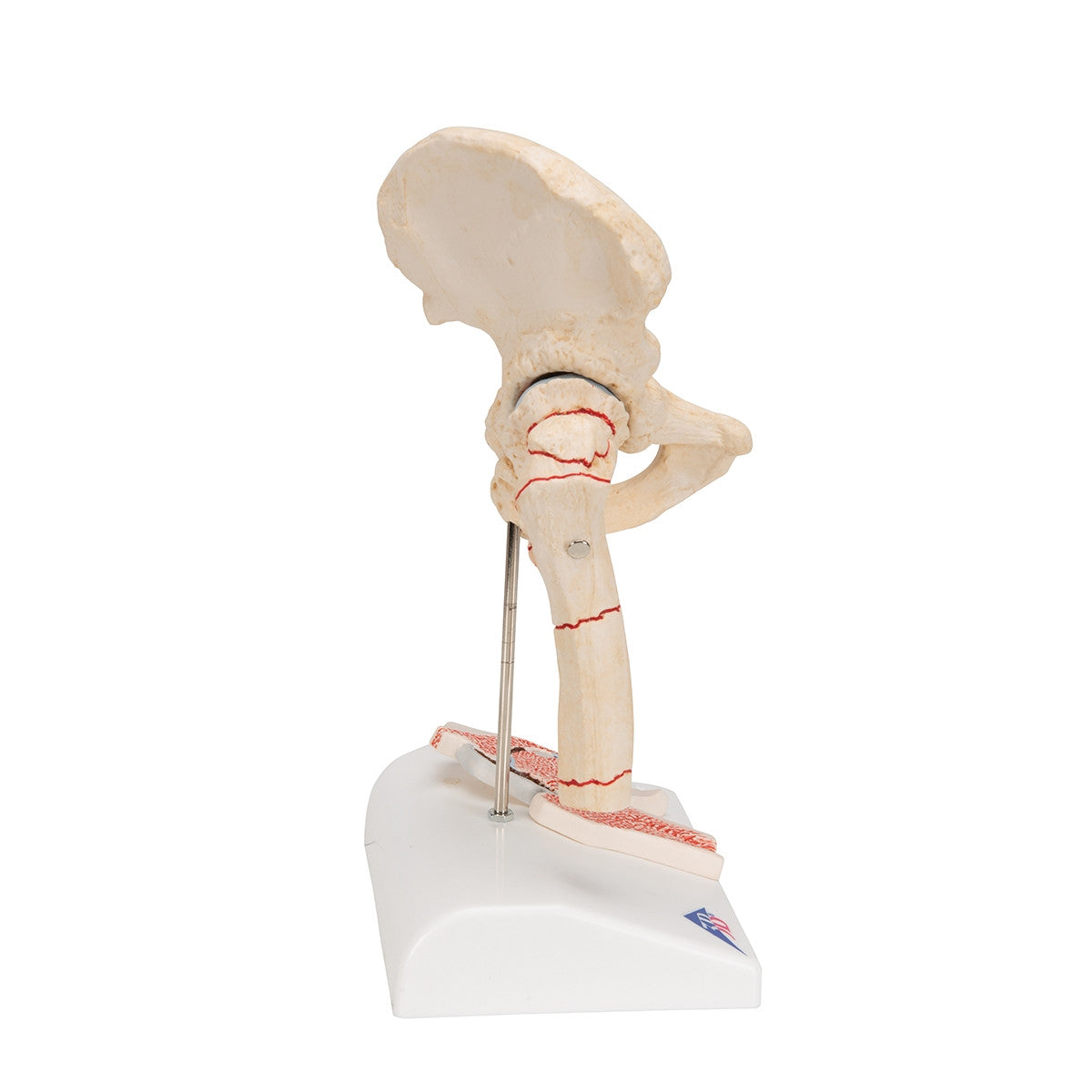
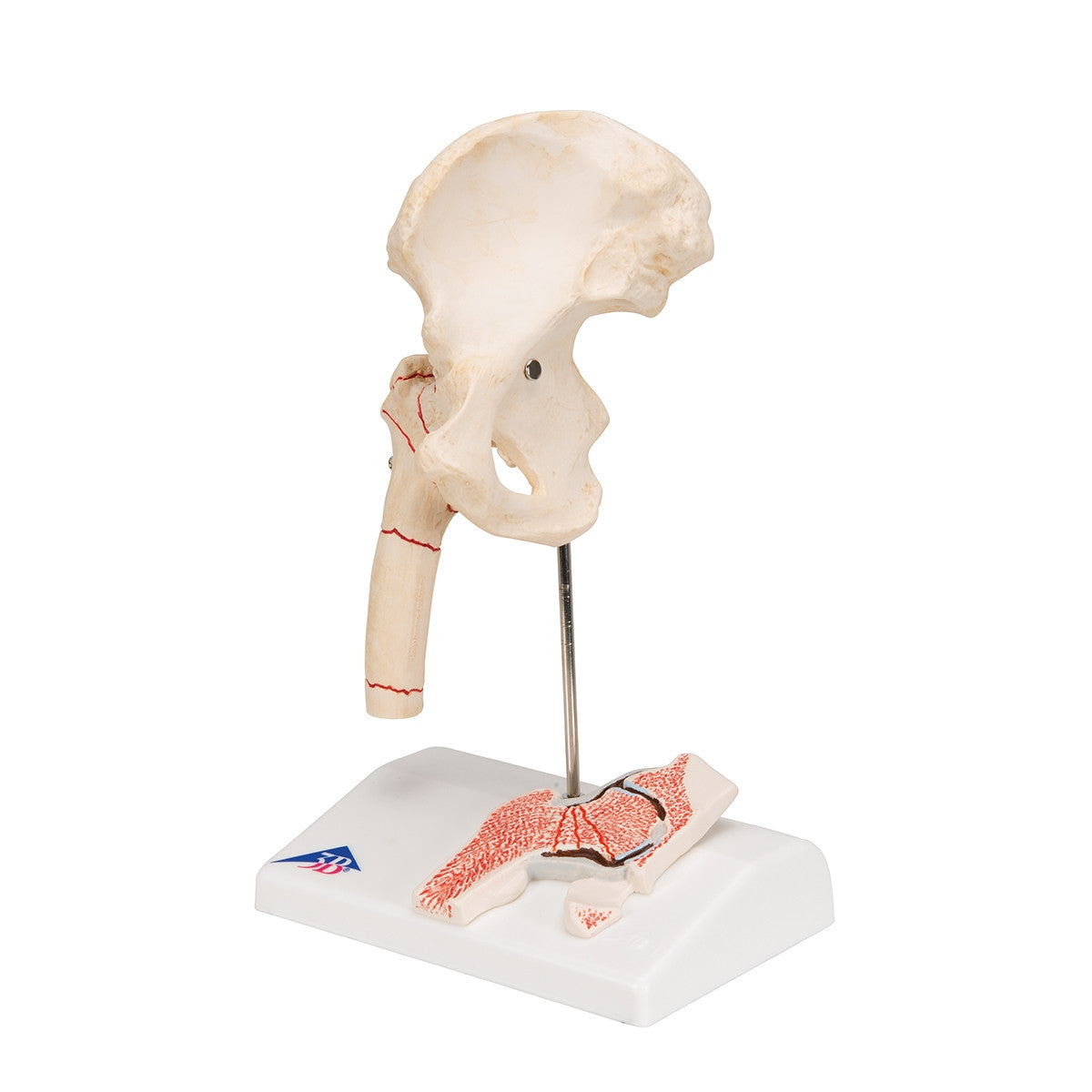
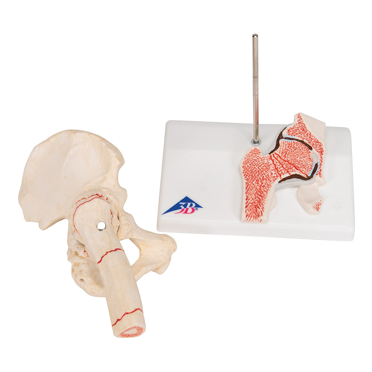
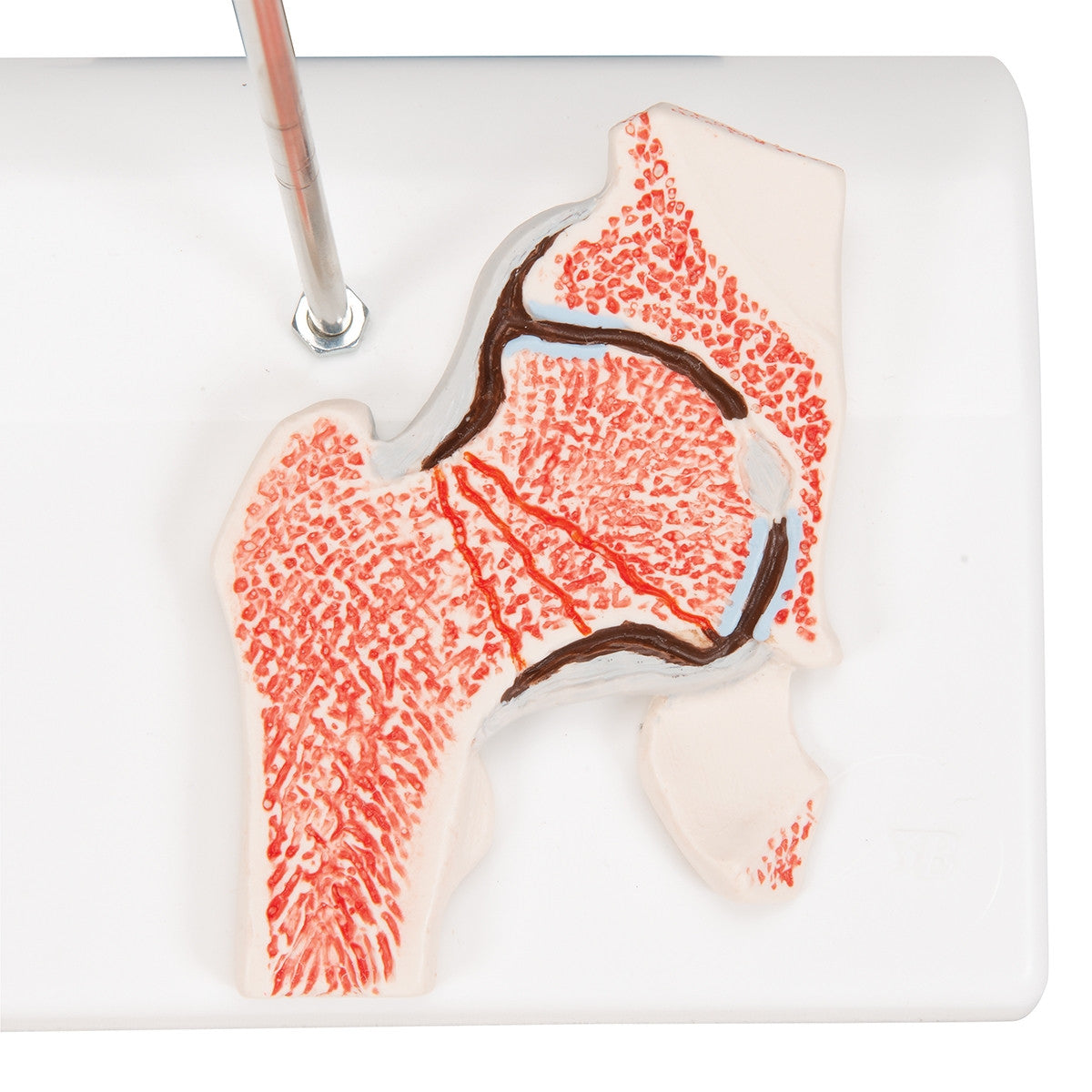
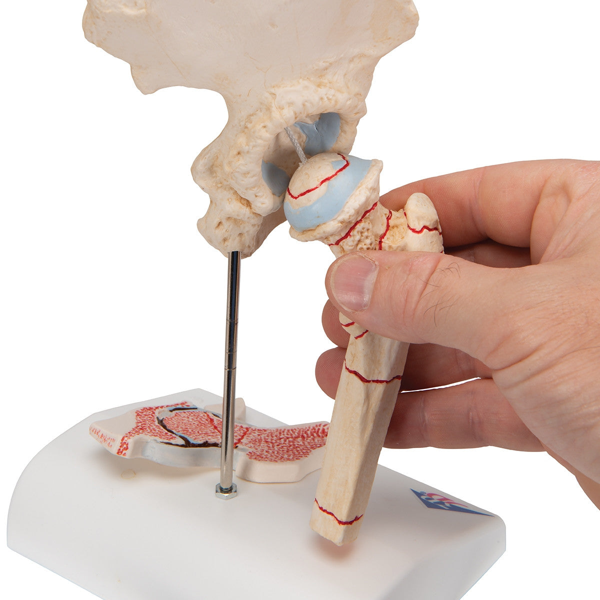
This femoral fracture model illustrates an elderly hip joint at half natural size. As well as the external anatomy of the hip joint, a frontal section through the femoral neck is also shown in relief on the base of the model. The hip joint model, A88 /1000175, shows the femoral fractures that occur most commonly in practice as well as wear and tear symptoms of the hip joint (coxarthrosis or hip osteoarthritis).
The hip fractures shown include:
- Medial femoral neck fracture
- Lateral femoral neck fracture
- Fracture through the trochanteric region (pertrochanteric femoral fracture)
- Fracture below the trochanters (subtrochanteric femoral fracture)
- Femoral shaft fracture
- Femoral head fracture
- Fracture of the greater trochanter
- Fracture or avulsion of the lesser trochanter (avulsion fracture of the lesser trochanter)
- Femoral fracture and hip osteoarthritis mounted on base.
This hip fracture model is ideal for orthopaedic departments, physio and GP clinics.
Download Femoral Fracture and Hip Osteoarthritis Model A88 /1000175 product manual here.
This model comes with 3B Scientific 3B Smart Anatomy app included for FREE. This features access to an anatomy course, including 3 digital anatomy lectures, 117 different virtual anatomy models and 39 anatomy quizzes. It also offers a FREE warranty upgrade from 3 to 5 years with every product registration. To unlock these benefits, scan the label located on your 3B Scientific anatomy model and register online.