


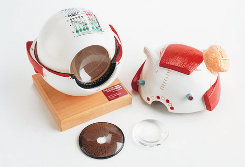
The Giant Eye Model is enlarged to six times its actual size and is a robust vinyl plastic replica that offers a wealth of valuable educational features.
On the outer surface of the eyeball, you can observe the cornea, providing a view of the iris and pupil, the prominent lacrimal (tear) gland, points of attachment for all six external eye muscles, the roots of the optic nerve, and the surrounding network of blood vessels.
The eyeball can be split in half, allowing for an in-depth examination of the interior, which includes the choroid layer of the retina. Within this section, various removable components are present, such as the vitreous ball, iris/cornea, and a Lucite lens that can magnify and invert images when taken out of the model. The retinal microstructures, including rods, cones, and others, are depicted in a highly detailed diagrammatic cross-section.
The Giant Eye Model has a total of 42 distinct features which are explained in an accompanying key.