


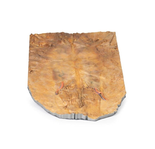
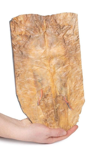
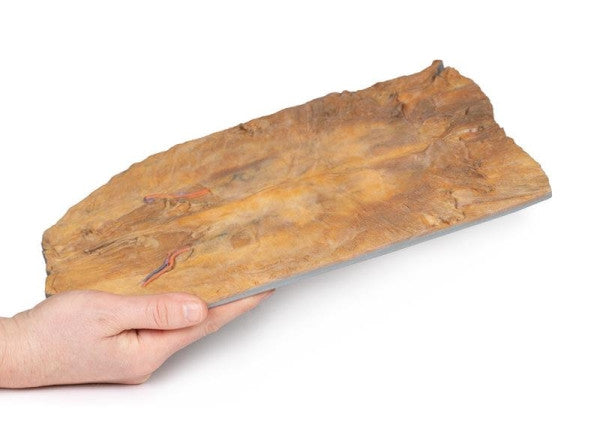
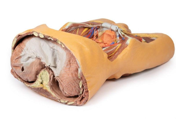
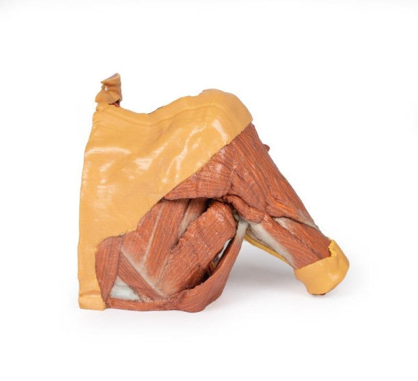
Internal Abdominal Wall
This 3D model reveals the internal surface of the anterior abdominal wall, exposing the convergence of muscle fibres, the arcuate line shift in aponeurosis orientation, and the path of the inferior epigastric arteries. In the midline, the median abdominal ligament covers the urachus.
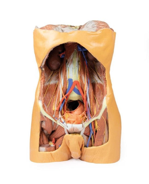
Posterior Abdominal Wall
This expansive 3D printed model exhibits the complete posterior abdominal wall in males, spanning from the diaphragm to the pelvic brim, encompassing pelvic structures and extending to the proximal thigh.
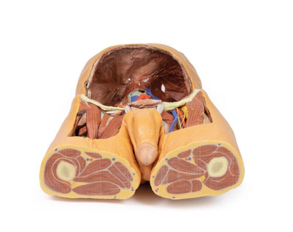
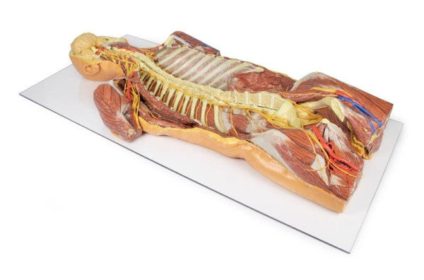
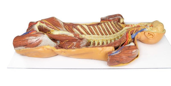
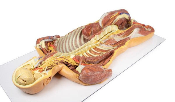
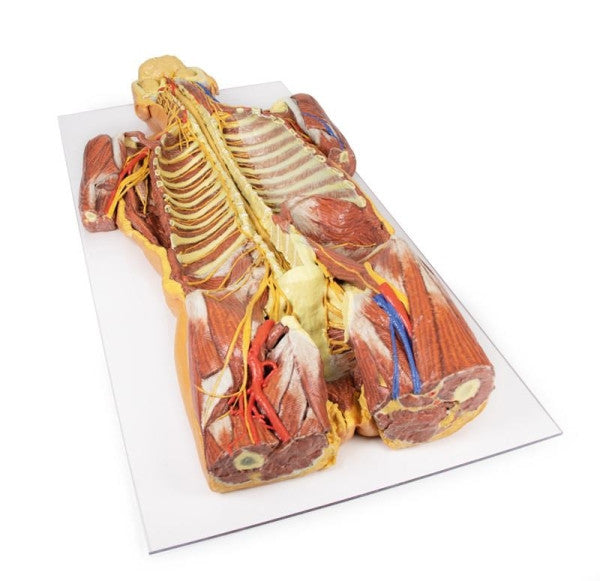
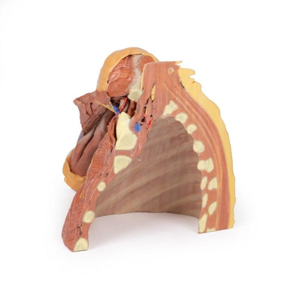
Posterior Body Wall (Ventral Deep Dissection)
This 3D-printed model offers a detailed ventral deep dissection of axial anatomy, covering the head, neck, axillae, thorax, abdomen, and proximal thighs. The removal of the anterior portions of the cranium and vertebral bodies from the cervical to the 5th lumbar region allows for a continuous view of the central nervous system structures and the origin of the segmental nerves in relation to other axillary and appendicular structures.
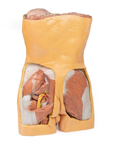
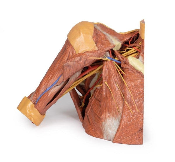
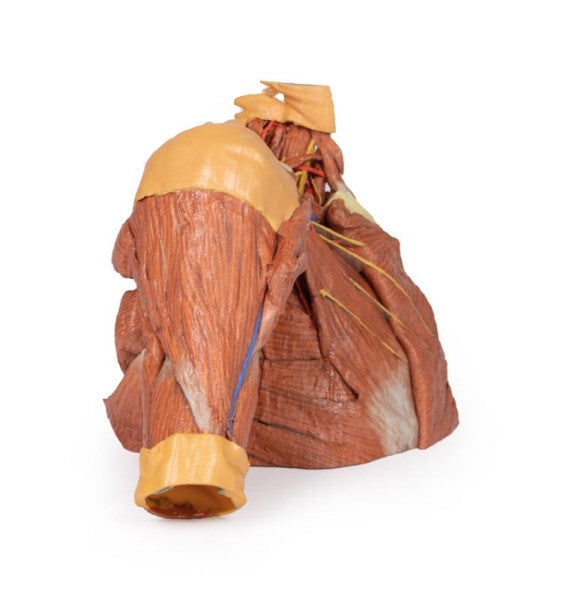
Right Thoracic Wall (Axilla and the Root of the Neck)
This 3D printed model reveals a parasagittal dissection of the right thoracic wall, axilla, and neck root, showcasing chest wall structures, the pectoralis minor's role in dividing the axillary artery, and the brachial plexus from C5-T1 roots to axillary exit.