


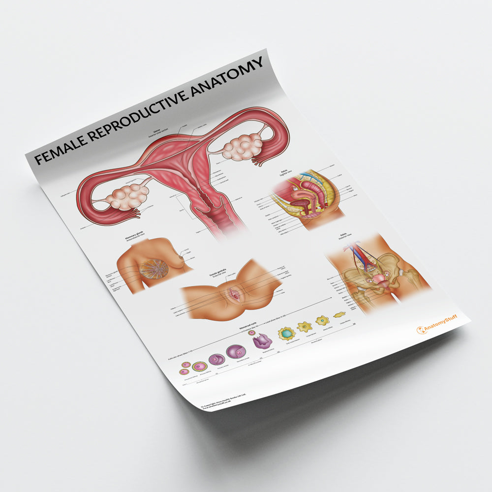
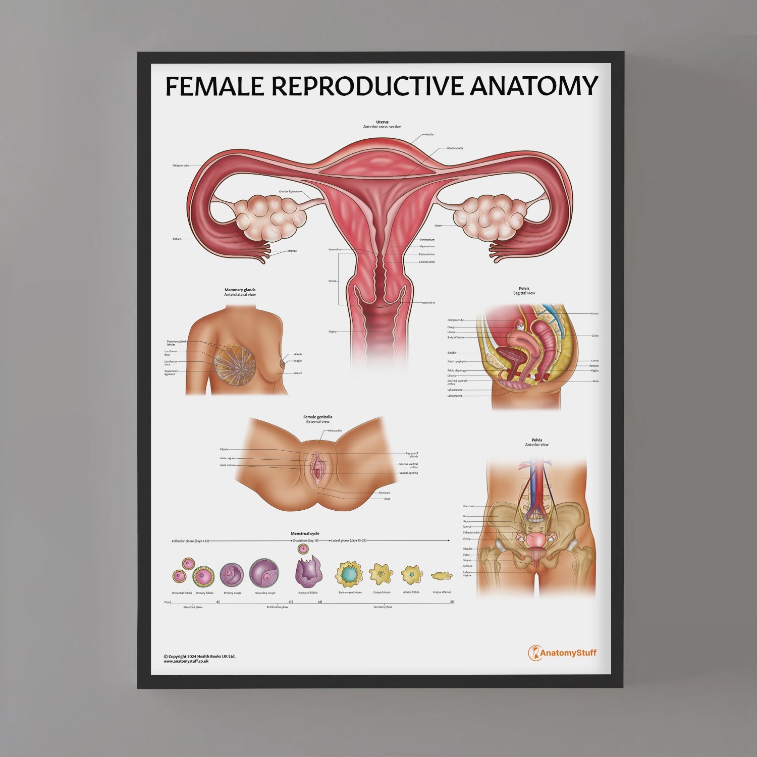
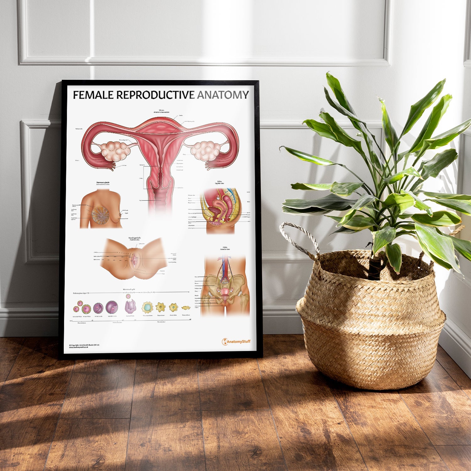
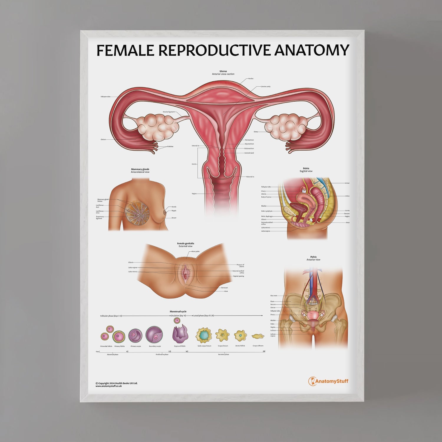

This brightly illustrated reproductive system chart details the anatomical features of the female reproductive organs. It would be ideal as a teaching aid for schools to university students, or as a patient educational aid in a medical clinic. Designed by a professional medical illustrator and exclusive to AnatomyStuff, our Female Reproductive System Anatomy poster shows the following anatomical details:
- Anterior view of the female body highlighting the position of the mammary glands and uterus
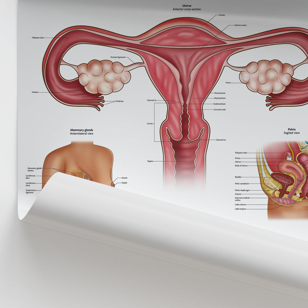
- Detailed anterior / cross-section view of the uterus, fallopian tubes and ovaries identifying 16 features including the cervix, layers of the uterine lining and associated ligaments
- Anterior view / cross-section of a mammary gland identifying the nipple, areola, lactiferous glands and ducts and fat layer
- Detailed illustration of an ovum (unfertilised egg), identifying the cell components including the chromosomes in the ooplasm and the cell membrane
- Lateral cross-section of the lower torso labelling reproductive organs including the cervix, uterus and features of the vagina
- Dorsal view of the external female genitalia identifying the different parts of the vagina
- Detailed view of a fallopian tube and ovary with a developing ovum and a summary and a schematic diagram of a typical 28 day menstrual cycle
Our collection offers a range of display options to meet your needs:
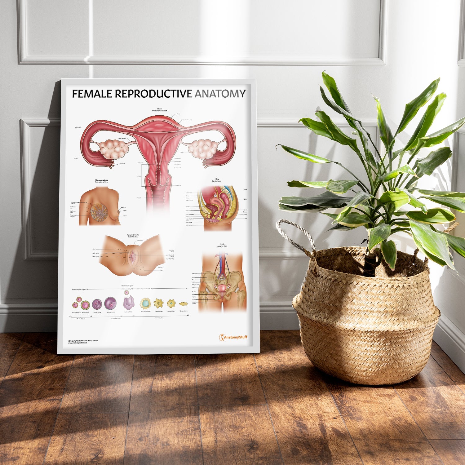
- Laminated Poster (50 x 65 cm): Ideal for clinic displays, featuring clearly labelled anatomical details. The durable lamination allows for annotation with washable markers (not supplied).
- Classic Semi-Glossy Prints: 45 x 60 cm, 60 x 80 cm, and 70 x 100 cm sizes. The semi-glossy finish enhances colours with a subtle shine, adding vibrancy to any setting.
- Framed Prints: Offered in black or white frames and sizes 45 x 60 cm, 60 x 80 cm, and 70 x 100 cm. Features 170 gsm matte paper with a smooth, non-reflective finish. Ready-to-hang with a durable pine wood frame.
