


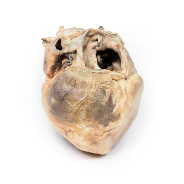

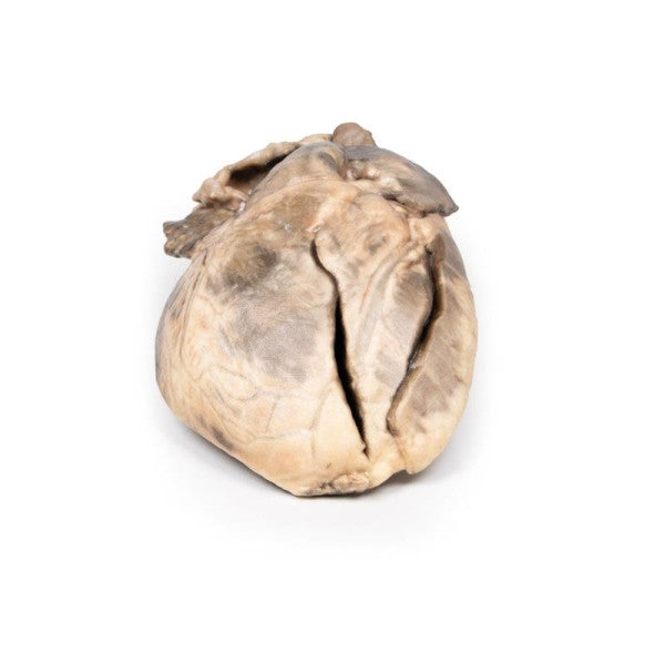
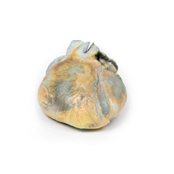
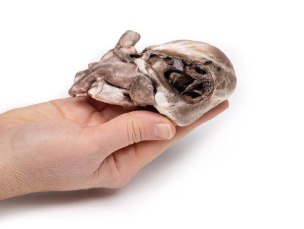
Atrial Septal Defect
The heart reveals a large oval-shaped defect in the inter-atrial septum, with only a small remaining crescentic rim. The left ventricle is small, and the right ventricle is hypertrophied. The pulmonary artery is significantly enlarged, and the cut edge below the pulmonary artery represents the left auricular appendage with an 8mm diameter lumen.
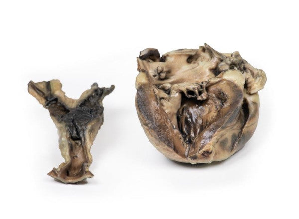
Hydatid Disease Affecting the Heart and Aorta
The specimens feature the heart with left ventricle exposure and the aorta showing atheromatous deposits. A large antemortem clot is present at the iliac bifurcation, extending into both common iliac arteries. The heart exhibits left ventricular wall hypertrophy and abnormal communication between the left ventricle and atrium, surrounded by thickened fibrous tissue. Hydatid cysts are found in the abdominal aorta and the channel connecting the left ventricle and left atrium.
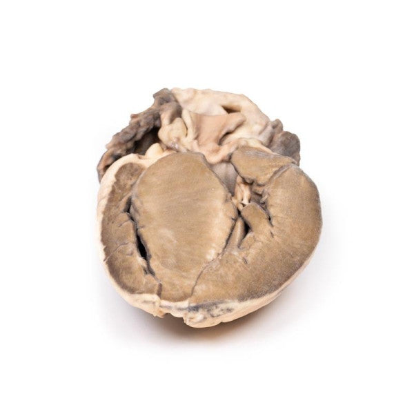
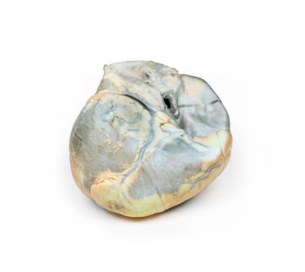
Hypertrophic Subaortic Stenosis
This is a heart section revealing the left and right ventricles and the interventricular septum. The notable anomaly is a significantly thickened interventricular septum and left ventricular hypertrophy. The enlarged ventricular septum encroaches on the left ventricle lumen.
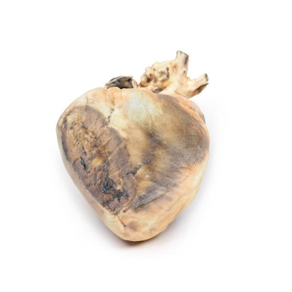
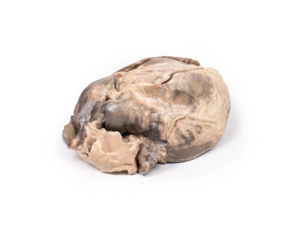
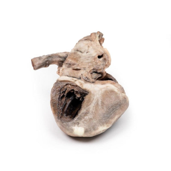
Right Ventricular Hypertrophy
The specimen displays a noticeably enlarged and hypertrophied right ventricle. Apart from this, everything else appears normal. This case illustrates right ventricular hypertrophy in a patient with emphysema.
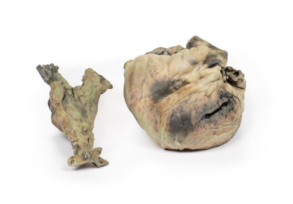
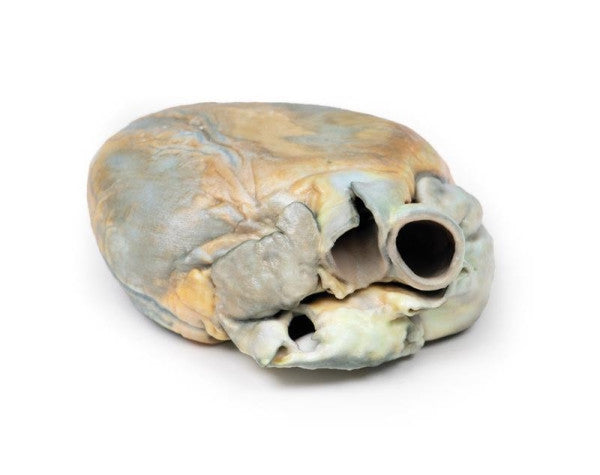
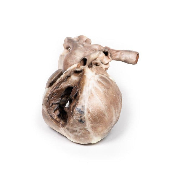
Tetralogy of Fallot
The child's heart exhibits significant right ventricular hypertrophy, a narrowed pulmonary outflow tract with a small, stenosed bicuspid pulmonary valve, and endocardial fibrosis. Probing reveals communication between the right ventricle and aorta, and from the pulmonary trunk to the dilated left pulmonary artery and descending aorta. The posterior view indicates an 8mm atrial septal defect at the foramen ovale, and a smaller 3mm atrial septal defect, with the left ventricle wall slightly thinner than the right ventricle.