


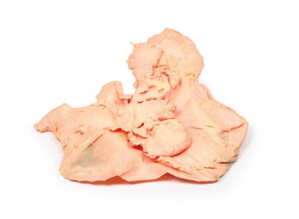
Adenocarcinoma of the Stomach
This post-mortem specimen shows a large 7 cm x 5 cm ulcer on the stomach's lesser curve, displaying shallow, broad features with raised edges and necrotic debris. Gastric rugae loss extends from the ulcer, and dissection exposes elevated edges with pale homogeneous tumour tissue. Two eroded arteries with the ulcer crater suggest a recent haemorrhage, and the pancreas adheres to the ulcer's serosal aspect.
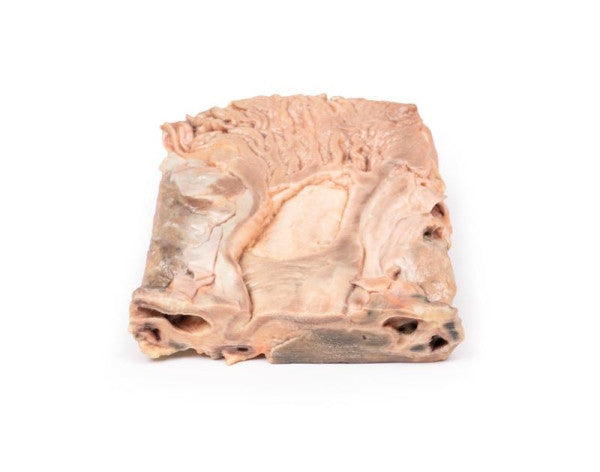
Chronic Gastric Ulcer
The 2 cm coronal tissue slice reveals a large oval-shaped ulcer with elevated borders near the gastro-oesophageal junction. The clean, smooth base lacks haemorrhage, while induration and fibrosis extend from the ulcer, causing radial convergence of gastric mucosal folds.
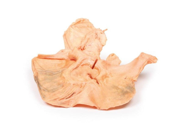
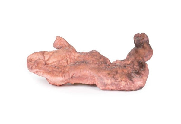
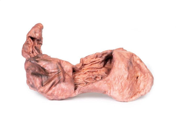
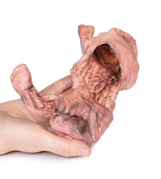
Stomach
This 3D model features a stomach with two dissection windows revealing the rugae and pylorus. The cardiac region and a portion of the proximal duodenum are preserved. The large window offers a clear view of the fundus and well-developed rugae, while the smaller window at the pyloric region highlights the thickening of the organ wall near the pyloric sphincter, just before the duodenum begins.
Stomach 3D Printed Anatomy Model - MP2080-76-1134
Dimensions: 25.5 cm x 35.5 cm x 14.5 cm
Weight: 0.5 kgs
Manufacturer: Erler Zimmer
Made in Germany